
- 2000 – completed DubnoMedicalTrainingCollege, speciality – medical assistant.
- 2005 – StomatologyAcademy of Ukraine. Qualification – Doctor of Stomatology.
- 2007 – completed internship on general dentistry based on Stomatology Academy of Ukraine. Specialty – Doctor of Stomatology.
- 2005 – participation in international lecture hall “Modern technology of treatment and preventative measures in practical stomatology” based on Scientific and Practical Center “Stomatology” (30 study hours). Participation in lecture “The way of real reflection of reality. Stratification methods Lorenzo Vanini”. Club «ENAMEL PLUS». Participation in lecture from course “Usage of material Filtek Supreme XT in clinical ctomatology”.
- 2006 – participation in VII All Ukrainian Seminar “The way into the world of mastership”. Study on course of thematic improvement “Usage of light curing technologies in teeth restoration”.
- 2008 – participation in lecture “Recure of root-canals. General consequences of imperfect treating of root-canals”. Master class, participation in the work of the work of scientific practical seminar “Whitening of teeth. Modern tendentions and approaches”.
- 2009 – participant of the enlarged meeting of Poltava regional group of Ukrainian Association of Doctors of Periodentistry.
- 2010 – participant in conference on restoration methods “Perfectibility in restoration methods. Restoration of teeth after endodontal treatment” of Dr. Andre Reis. Brazil.
- 2011 – participant in the conference “Paradontium. Deceases and treatment”. Participant in the seminar “Usage of whitening toothpastes. Influence of abrasiveness to paradontium and tooth enamel”.
- From 2012 – till present time is a dentist of the surgery department of «Oxford Medical» clinic.
Dentistry
Dentistry
- Dental implants
- Veneers
- Sedation Dentistry
- Teeth whitening
- Teeth treatment
- Microscopic dentistry
- Dental disease prevention
- Tooth decay treatment
- Tooth extraction
- WISDOM TOOTH REMOVAL
- Dentist consultation
- Endodontics – root canal treatment
- Gums treatment
- Treatment of parodontitis
- Emergency dental care
- Pulpitis treatment
- Apical periodontitis treatment
- Treatment of Dental Cyst
- AIR FLOW TEETH CLEANING
- Dentures
- Diagnosis
- Aesthetic dentistry
- Orthodontics

Have you ever experienced the consequences of improperly diagnosed disease? Even if no, you have probably heard from friends about dental problems, which remained unnoticed, developed and led to tooth extraction. The diagnostic error impedes any efforts to cure teeth. That is why it is very important to find the clinic, which has modern diagnostic equipment and experienced dentists, capable of making correct diagnosis.
DIAGNOSTIC METHODS
The Dentistry Department of Oxford Medical clinic uses the following advanced diagnostic methods:
- X-ray imaging;
- panoramic X-ray imaging;
- teleradiology;
- CT scan;
- diagnosis in articulator;
- Schulz’s method;
- T-Scan III occlusal analysis;
- palpation of temporomandibular joint and masticatory muscles of TMJ;
- diagnosis with intraoral camera.
Forget about the old-fashioned film X-ray, which state and most private clinics use – it gives little information due to the absence of possibility of enhancing the imprint on the film. We use the X-ray scan, which allows making a precise digital X-ray image of your tooth and immediately display it on the screen. During the consultation, the doctor enhances the image for identifying smallest defects and offers you different treatment options.
The use of panoramic X-ray imaging allows obtaining a panoramic image of the upper and lower jaw. When displayed enhanced on the computer, it enables to perform the comprehensive examination of the whole oral cavity.
The panoramic X-ray imaging system in Oxford Medical clinic is additionally fitted with a device for making a front and profile image of the skull – cephalostat of the last model. The obtained teleradiology fully displays the condition of joints and bones of both jaws; that is why orthodontists regularly use it while building the orthodontic models.
The above-listed methods relate to 2D diagnosis, which provides a significant amount of information; however, it often requires detailing, using 3D diagnostics. Only several clinics of the country have the digital panoramic X-ray scan, which allows making 3D images of teeth, bone tissue, temporomandibular joint, and even the motion computed tomography of the joint, and Oxford Medical is among them.
During the denture placement, the diagnosis and preliminary preparation for the procedure is performed in the individualized SAM-articulator, which allows creating an accurate model of your jaw and precisely copy its movement. This approach allows making all manipulations so that they do not impact your teeth. For example, no odd “elevations” will appear, and the teeth will not be chipped, the crowns – get cracked etc.
The aesthetic aspect is also important for performance of dental manipulations. Thanks to Schulz’s method, the doctor demonstrates a future external view of teeth before starting their treatment. This method of wax modelling enables the dentist to make corrections before the beginning of a treatment and makes sure that the final result will fully satisfy the patient.
T-Scan III occlusal diagnosis guarantees the preservation of your teeth after treatment. The essence of the diagnosis consists in the insertion of electronic sensors inside the oral cavity, placed on the thin plate. After that, all imperfections are displayed on the monitor, and then the teeth are filed down according to much more precise data, than after the copy paper, which was used for long in Soviet dentistry.
In 3 months, the dentist will repeat the T-scan procedure (occlusal analysis), because the muscle load slightly changes after placement of any types of dentures, and the change of their tone can provoke unacceptable contacts. The man gets used, but such contacts can lead to destructive process; that is why this procedure is very significant. The T-rescanning is performed in Oxford Medical free of charge.
The highly qualified doctors of Oxford Medical also competently perform one of the underlying methods of diagnosis – palpation of temporomandibular joint and masticatory muscles that is essential for correct diagnosis.
One more innovation is the alternative to a miniature mirror, which was previously inserted inside the oral cavity – the intraoral camera, which allows performing a maximally precise examination and noticing insignificant negative changes. Thanks to this method, you will not just hear the doctor’s opinion, but you will see the real condition of your teeth on the plasma screen.
WHY OXFORD MEDICAL?
The Oxford Medical uses the last models of high quality equipment, while the highly qualified doctors perform all the manipulations, which regularly attend courses not only in Ukraine, but also abroad.
We guarantee the diagnosis precision. Trust professionals!
Our EXPERTS
all experts
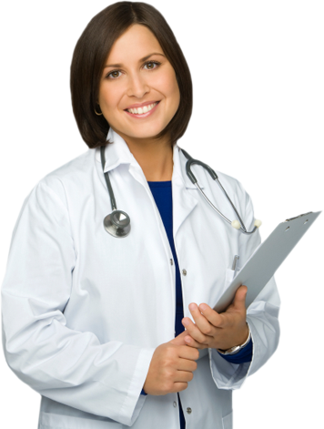
Berlinetz Irina Valerievna Dentist
information about doctor


reviews
Владислав Гончаренко 12-04-2024 Хочу порекомендувати лікаря-хірурга Якименко Р.О.Видаляв 3 зуба мудрості та встановив імплант, я дуже задоволений його роботою, все пройшло якісно і безболісно. Хоча трішки боявся, але все пройшло на ура, навіть не очікував, що після наркозу буде взагалі комфортно і не боляче!Всім рекомендую!!!
department: Dentistry
Єва 19-03-2024 Велика подяка Анжеліці Шахівні за професіоналізм, уважне ставлення і комплексний підхід! Лікар надала вичерпну консультацію, а чистка зубів, реставрація пройшла безболісно і комфортно, на найвищому рівні. Рекомендую клініку кожному, залишилась максимально задоволена якістю послуг!
department: Dentistry
Анатолий 14-03-2024 Хочу висловити подяку лікарю пародонтологу Лєсковій Олександрі. Професіонал своєї справи, все зробила дуже якісно, дала дієві поради за доглядом ротової порожнини. Задоволен тим, що звернувся саме до неї. Велике дякую!
department: Dentistry
Олена 17-02-2024 Велика подяка спеціалістам клініки(відділення Оболонь)!❤️Хірургу Маляренко Марині Євгенійовні: красива,молода,чуйна,ПРОФЕСІОНАЛ своєї справи.Я дуже задоволена:і підхід,і виконана робота,і результат❤️Асистент Тарас-Молодець:виконує свою роботу на"5"!! Менеджер Леся- робить перебування в клініці ще комфортнішим:Дякую!Адміністратор сьогодні була Марина: Дякую!!Рекомендую❤️Подяка кліниці за чудову КОМАНДУ!! Успіхів вам всім!!
department: Dentistry
Іванна 04-02-2024 Хочу подякувати Марині Євгенівні за чуйність, професіоналізм та уважне ставлення до пацієнта. Видалення зуба в ургентній ситуації пройшло максимально комфортно. Дуже красива і приємна лікар. Дякую!
department: Dentistry
Leave a recall
Special offers

current information


Video reviews
A true guarantee of quality and comfortable treatments are thousands of satisfied patients


Select city
Oxford Medical Clinic
Glubochitskaya street, 40X
phone:
+38 (044) 204-40-40
show - Viber, Telegram
Pavlovskaya street, 26/41
phone:
+38 (044) 204-40-40
show - Viber, Telegram
st. Predslavinskaya 29
phone:
+38 (044) 204-40-40
show - Viber, Telegram
Bereznyakovskaya street, 30B
phone:
+38 (044) 204-40-40
show - Viber, Telegram
Sribnokilskaya street, 20
phone:
+38 (044) 204-40-40
show - Viber, Telegram
st. Mokra, 20A
phone:
+38 (044) 204-40-40
show - Viber, Telegram
Ivana Kramskogo street, 9
phone:
+38 (044) 204-40-40
show - Viber, Telegram
Obolonska Naberezhna Street 1, building 2
phone:
+38 (044) 204-40-40
show - Viber, Telegram
st. Mikhaila Grishka, 1
phone:
+38 (044) 204-40-40
show - Viber, Telegram
Knyaziv Ostrozskykh street, 46/2
phone:
+38 (044) 204-40-40
+38 (097) 204-84-84 - Viber, Telegram
Mykhaila Dragomirova street, 14B
phone:
+38 (044) 204-40-40
show - Viber, Telegram
телефон:
+38 (044) 204-40-40
+38 (097) 204-84-84 - Viber, WhatsApp, Telegram
Irpen. Universitetskaya street, 2/1; building 3
phone:
+38 (044) 204-40-40
show - Viber, Telegram
make an appointment
If you can not find the desired information - contact us convenient for you