


Contents:
- What is choledocholithiasis?
- Causes of stone formation in the bile ducts
- Choledocholithiasis: symptoms of the disease
- Possible complications of choledocholithiasis
- Diagnosis of choledocholithiasis
- Treatment of choledocholithiasis
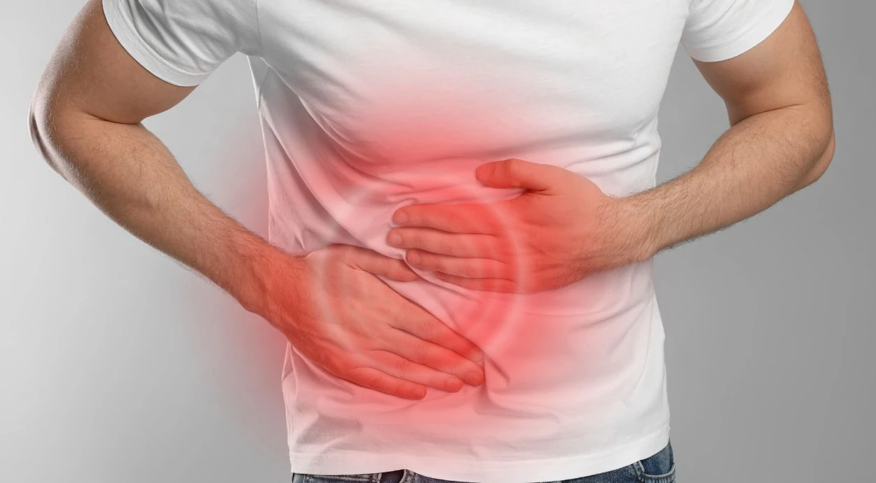
Pain in the right upper abdomen, nausea, and yellowing of the skin may occur when stones block the flow of bile from the liver to the intestine. This condition is called choledocholithiasis. It may not cause noticeable symptoms for a long time, but when the duct becomes blocked, there is a risk of acute inflammation and other dangerous complications. Therefore, it is important to see a doctor promptly if characteristic signs appear.
In this article, we will explain why stones form in the bile ducts, what symptoms accompany the disease, and why it is dangerous. We will also explain which examinations help establish the diagnosis and which methods are used to remove the stones.
What is choledocholithiasis?
Choledocholithiasis is the presence of stones in the common bile duct (choledochus), which connects the bile ducts of the liver and the gallbladder to the duodenum. Bile flows through it into the intestine, where it participates in the digestion of fats. Stones can vary in size and be located in any part of the common bile duct, partially or completely blocking its lumen.
Choledocholithiasis differs from gallstone disease in the location of the stones: in the first case, they are located in the common bile duct, while in the second, they are usually found in the gallbladder. These conditions may occur together, but stones in the common bile duct are sometimes also detected in patients who have previously had their gallbladder removed.
Causes of stone formation in the bile ducts
Most often, stones appear in the bile ducts as a result of their migration from the gallbladder. Much less frequently, stones form directly in the bile ducts.
The main causes of choledocholithiasis:
- Gallstone disease. Stones that have formed in the gallbladder can migrate through the cystic duct into the common bile duct. This is the most common mechanism by which the disease develops.
- Bile stasis. When the bile ducts narrow or their patency is impaired, bile is retained, creating conditions for its components to precipitate and form stones.
- Bile duct infections. Certain bacteria alter the composition of bile and contribute to the formation of pigment stones, particularly those composed of calcium bilirubinate.
According to the American Society for Gastrointestinal Endoscopy, stones in the common bile duct are simultaneously detected in 10–20% of patients with symptomatic gallstone disease. This confirms that the migration of stones from the gallbladder is an important mechanism in the development of choledocholithiasis.
Choledocholithiasis: symptoms of the disease
Choledocholithiasis may remain without noticeable manifestations for a long time. Symptoms usually occur when a stone partially or completely blocks the lumen of the common bile duct and disrupts bile flow. The severity of the manifestations depends on the degree of obstruction and its duration.
Stones in the bile ducts — symptoms:
- sudden, intense pain in the right upper abdomen or upper part of the abdomen, which may radiate to the back or under the right shoulder blade;
- nausea and vomiting, which do not always provide relief;
- yellowing of the skin and whites of the eyes due to increased bilirubin levels in the blood;
- darkening of the urine to the color of strong tea;
- light-colored or grayish stools due to insufficient bile pigments entering the intestine;
- itching of the skin, especially with prolonged impairment of bile flow.
The manifestations may be intermittent. If the stone shifts and the patency of the duct is temporarily restored, pain and other symptoms sometimes decrease or disappear. However, this does not mean that the problem has been completely resolved.
Doctors at Oxford Medical warn: the combination of pain in the right upper abdomen, jaundice, and fever with chills is particularly dangerous. These three signs are known as Charcot's triad and may indicate the development of acute cholangitis — an infectious inflammation of the bile ducts.
The nature of the pain and accompanying signs cannot reliably determine the presence of stones in the common bile duct. Similar complaints may occur with other diseases of the digestive organs, so an examination is necessary to establish the diagnosis.
Possible complications of choledocholithiasis
If a stone in the bile duct blocks its lumen, bile cannot flow normally into the intestine and accumulates in the bile ducts. Prolonged impairment of its flow can lead to liver damage and, if an infection develops, to acute inflammation. In addition, a stone can block the area where the bile duct joins the pancreatic duct and cause pancreatitis.
The main complications of the disease:
- obstructive jaundice — an increase in bilirubin levels in the blood due to impaired bile flow;
- acute cholangitis — bacterial inflammation of the bile ducts, which in severe cases can lead to sepsis;
- acute biliary pancreatitis — inflammation of the pancreas due to a stone blocking the outflow of its secretions in the area where the ducts share a common outlet;
- secondary biliary cirrhosis — gradual scarring of liver tissue resulting from prolonged obstruction of the bile ducts.
According to the National Center for Biotechnology Information (NCBI), bile duct obstruction, cholangitis, and pancreatitis are among the main dangerous consequences of choledocholithiasis. These conditions may require urgent restoration of bile flow.
The greatest threat is acute cholangitis with the development of sepsis, which may be accompanied by a sharp drop in blood pressure and impaired consciousness. If these signs appear, emergency medical services must be called immediately.
At the same time, severe complications do not occur in all patients. Timely detection and removal of stones help restore the patency of the bile ducts and reduce the risk of further organ damage.
Diagnosis of choledocholithiasis
.jpg)
Diagnosis begins with a consultation with a gastroenterologist. The doctor clarifies the nature of the pain, checks for jaundice, and determines whether the patient has previously had gallbladder diseases or surgery to remove it. To confirm the diagnosis, it is necessary to determine whether there are stones in the common bile duct, exactly where they are located, and how severely bile flow is impaired.
The main examination methods:
- Laboratory blood tests — measurement of bilirubin, ALT, AST, alkaline phosphatase, and gamma-glutamyltransferase levels to assess liver function and detect signs of impaired bile flow. If necessary, inflammatory markers and lipase levels are checked to rule out pancreatitis.
- Abdominal ultrasound — makes it possible to detect dilation of the bile ducts and assess the condition of the liver and gallbladder. Large stones are sometimes directly visible, but small stones may remain undetected.
- Magnetic resonance cholangiopancreatography (MRCP) — an examination that makes it possible to obtain detailed images of the bile ducts and pancreatic ducts without inserting an endoscope. It helps determine the location and size of stones.
- Endoscopic retrograde cholangiopancreatography (ERCP) — a procedure during which a contrast agent is introduced into the bile ducts through an endoscope and X-ray images are taken. It is mainly used when there is a high probability of stones being present and they need to be removed immediately.
The examination makes it possible to establish an accurate diagnosis and select appropriate treatment.
Treatment of choledocholithiasis
The main goal of treating choledocholithiasis is to remove the stones and restore the free flow of bile into the duodenum. Most often, an endoscopic procedure is used for this purpose, allowing stones to be extracted without incisions in the abdominal wall. The choice of method depends on the size and number of stones, their location, the patency of the bile ducts, and the presence of complications.
The main treatment methods:
- Endoscopic stone removal (ERCP). An endoscope is inserted through the mouth into the duodenum, after which the doctor gains access to the common bile duct. If necessary, its opening is widened, and the stones are removed using a special basket or balloon.
- Lithotripsy. If a stone is too large or cannot be removed whole, special fragmentation methods are used. After the stone is broken up, its fragments are extracted from the duct.
- Bile duct stenting. If it is impossible to remove all the stones during the first procedure, the doctor may temporarily place a stent to ensure bile drainage. A repeat procedure is performed later.
- Surgical treatment. If endoscopic methods are ineffective or technically impossible, the stones are removed during surgery. The procedure may be performed laparoscopically or through open surgery, depending on the clinical situation.
According to the recommendations of the European Society of Gastrointestinal Endoscopy (ESGE), confirmed stones in the common bile duct are recommended for removal even in the absence of symptoms, provided that the patient's condition allows the procedure to be performed.
After the common bile duct has been cleared, patients who still have a gallbladder containing stones are usually advised to have it removed — a procedure called cholecystectomy. This helps prevent stones from entering the ducts again.
Doctors at Oxford Medical emphasize: taking choleretic medications independently does not reliably eliminate stones and may be dangerous if the duct is blocked. Timely restoration of bile flow and elimination of the source of stone formation help reduce the risk of recurrent attacks and severe complications.
Choledocholithiasis requires timely diagnosis and treatment, even if the symptoms decrease or temporarily disappear. Removing the stones and restoring bile flow help prevent dangerous complications and preserve the health of the biliary system.
The information in this article is provided for informational purposes and is not intended as instructions for self-diagnosis or self-treatment. If symptoms of a disease appear, you should consult a doctor.
Sources: