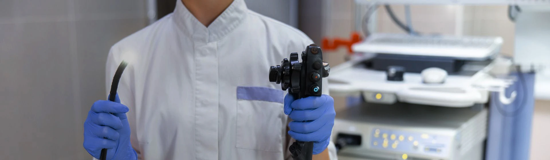
Chromoendoscopy is performed during gastroscopy or colonoscopy. First, the doctor performs a standard examination of the mucous membrane, after which, if necessary, proceeds to chromoendoscopy.
In classic chromoendoscopy, a special dye is applied to the mucosa through the endoscope. After it is distributed, the doctor re-examines the required area and assesses it under magnification on the screen.
Virtual chromoscopy is performed without applying dyes. Special endoscopic imaging modes are used for it, which enhance the contrast of the vascular pattern and the structure of the mucous membrane.
If a polyp, an area of dysplasia, an ulcer with atypical changes, or another formation is detected during the examination, the doctor may perform a targeted biopsy. A small tissue fragment is taken through the instrumental channel of the endoscope and sent for histological examination, which makes it possible to determine the nature of cellular changes and confirm or exclude a malignant process.