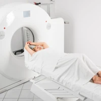
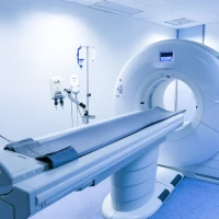
During the examination, the patient lies down on the table of the computed tomography scanner, and the leg is positioned in the position necessary for scanning. To obtain high-quality images, it is important to remain still throughout the entire procedure.
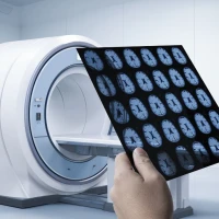
During the operation of the tomograph, a series of layer-by-layer images is performed, on the basis of which detailed images of the bones and articular surfaces are formed. This allows the doctor to assess the nature of the changes and clarify the diagnosis.
Scanning usually lasts only a few minutes and does not cause pain or significant discomfort. After the procedure is completed, the radiologist analyzes the obtained data and prepares a written conclusion.
With the CT results, it is recommended to consult the doctor who referred for the examination. This may be an orthopedic traumatologist, rheumatologist or another specialized specialist who will determine the further treatment tactics.