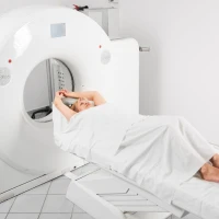
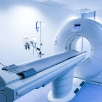
During the study, the patient is positioned on the table of the computed tomography scanner, and the arm is fixed in the position necessary for scanning. It is important to remain still throughout the procedure in order to obtain the clearest possible images.
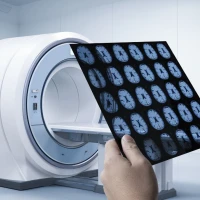
The tomograph performs a series of layer-by-layer images of the elbow area at different angles. Based on them, detailed images are formed, which allow assessment of the condition of bone structures and detection of pathological changes.
Scanning usually lasts only a few minutes and is not accompanied by pain or discomfort. After the study is completed, the radiologist analyzes the obtained data and prepares a written conclusion.
Based on the CT results, the patient can consult a specialized specialist to determine further treatment tactics. Most often this is an orthopedic traumatologist, who assesses the results of the study, clarifies the diagnosis and, if necessary, prescribes further treatment.