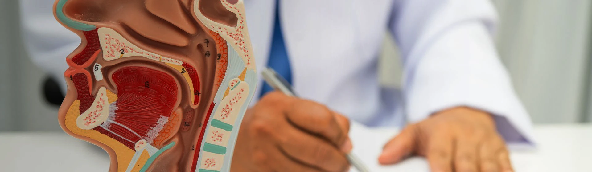
Rhinoscopy is usually performed during an appointment with an otolaryngologist. First, the doctor clarifies the patient’s complaints, after which they perform endoscopic diagnostics of the nasal cavity and nasopharynx.
A thin endoscope with a camera and light source is used for the examination. The doctor carefully inserts it into the nasal passage and sequentially examines the mucous membrane, nasal septum, nasal passages, nasal turbinates, and, if necessary, the nasopharynx. The image is displayed on a monitor in magnified form, which makes it possible to assess the condition of the tissues in detail.
During the procedure, the patient may feel slight discomfort or a brief urge to sneeze, but there is usually no pronounced pain. If necessary, before the examination the doctor may treat the mucous membrane with a local anesthetic and, to reduce swelling, use a vasoconstrictor medication.
Rhinoscopy lasts approximately 5–10 minutes. After it is completed, the doctor explains the results and, if necessary, recommends further examination or treatment.