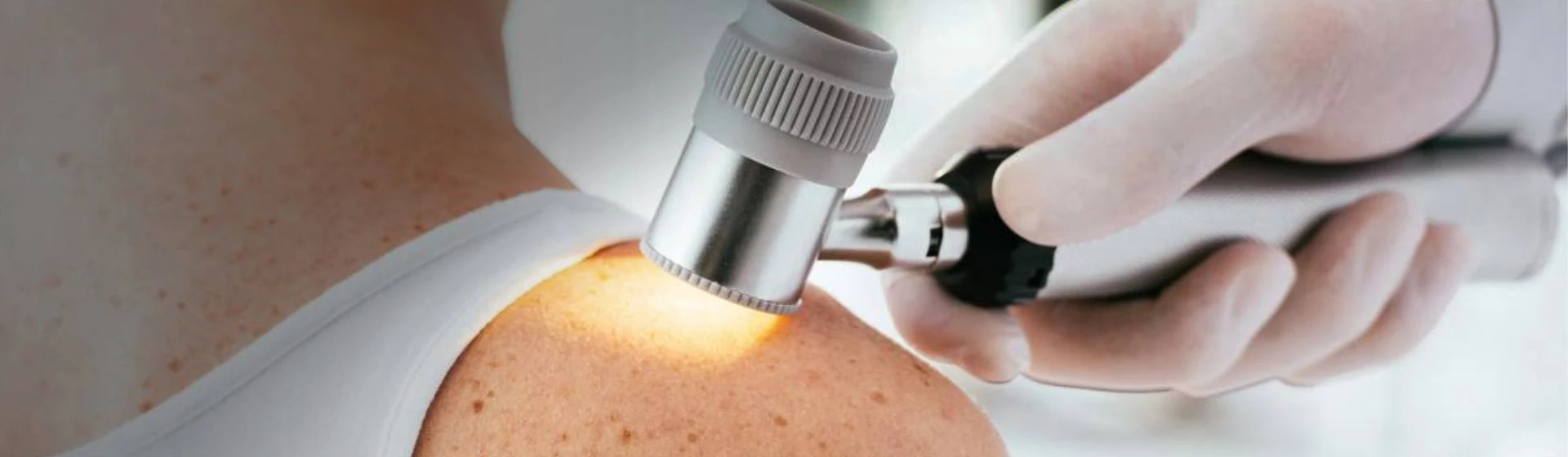
Dermatoscopy is a non-invasive diagnostic method during which a doctor uses a special optical device to examine moles and other skin neoplasms in detail. Magnification and special lighting make it possible to see pigment and vascular structures that are invisible to the naked eye. Dermatoscopy of a nevus helps identify signs resembling melanoma and other malignant skin tumors and determine further management.
Depending on the method of obtaining the image, two types of examination are distinguished:
- Analog dermatoscopy — the doctor examines the neoplasm directly through an optical dermatoscope.
- Digital dermatoscopy — the image of the mole is photographed and stored in digital format for subsequent comparison and monitoring of changes.
At the Oxford Medical medical center, a German-made HEINE DELTA 20 Plus dermatoscope is used. Thanks to tenfold magnification, LED lighting, and a polarizing filter, the doctor can examine the structure of the neoplasm, the characteristics of pigment distribution, and the vascular pattern in detail. If necessary, digital photographic documentation is performed to save the image for further observation and determine whether it is necessary to remove the mole.
Doctors at Oxford Medical note: skin dermatoscopy helps detect suspicious changes but does not replace histological examination. If the doctor suspects a malignant process, a biopsy followed by histological examination of the tissue may be necessary to establish the final diagnosis.